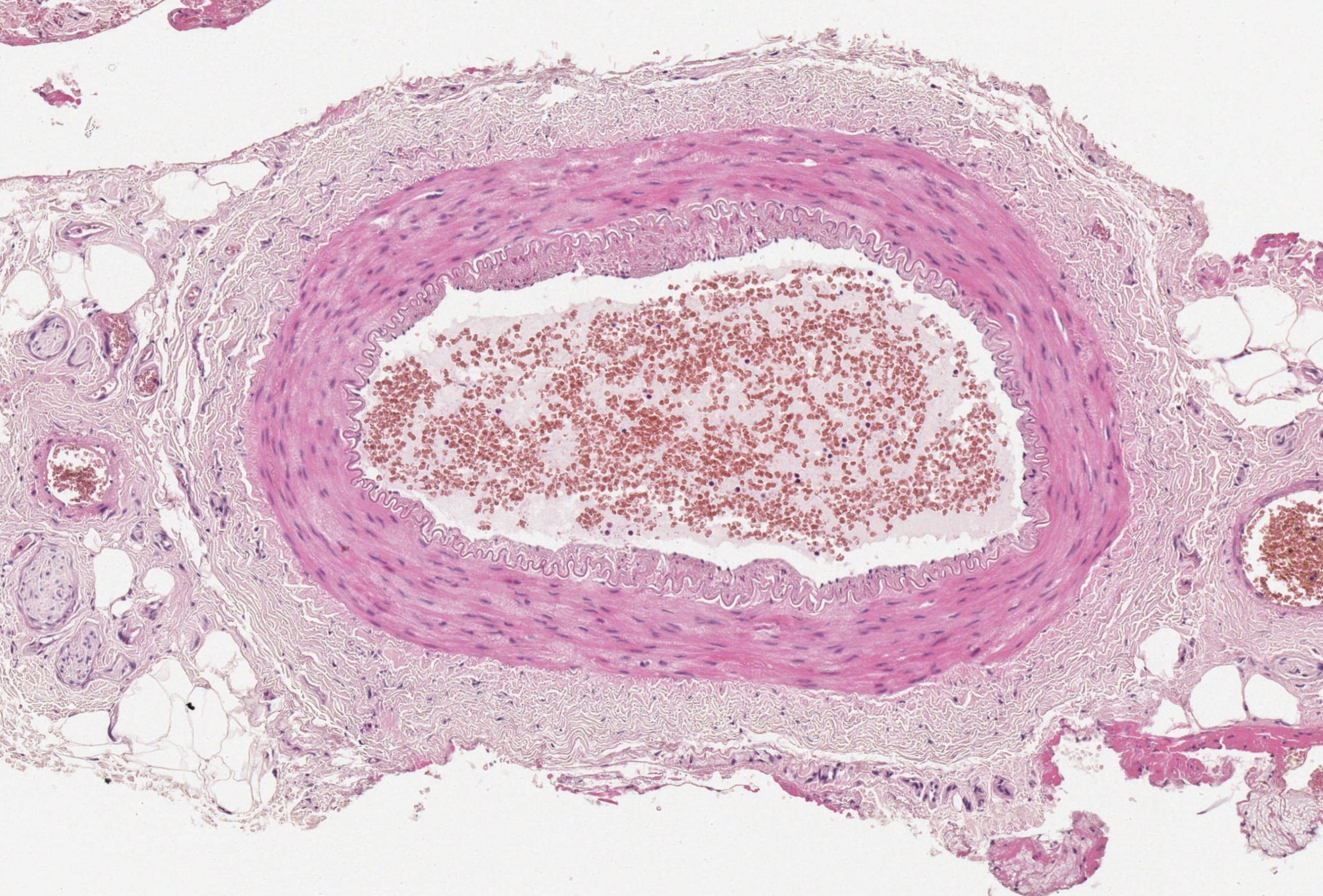
Normal temporal artery histology
A normal temporal artery comprises 3 distinct layers. These are: Tunica intima Tunica media Adventitia Each structure has its unique properties which can be summarised
This section explores the normal temporal artery histology as well as the differences between multinucleated giant cells and granulomatous inflammation. In addition, examples of the four main patterns of GCA, described by Hernández-Rodríguez et al, are explored.
The four patterns of GCA are as follows:

A normal temporal artery comprises 3 distinct layers. These are: Tunica intima Tunica media Adventitia Each structure has its unique properties which can be summarised
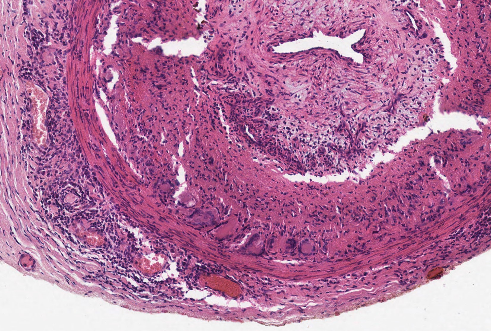
Multinucleated giant cells/ giant cells are often used interchangeably and refer to large cells with multiple nuclei.
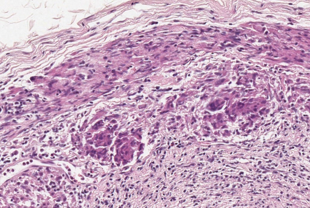
A granuloma is defined as collections of mononuclear cells (lymphocytes and macrophages, with or without the formation of giant cells. Granuloma/ granulomatous inflammation example Multinucleated
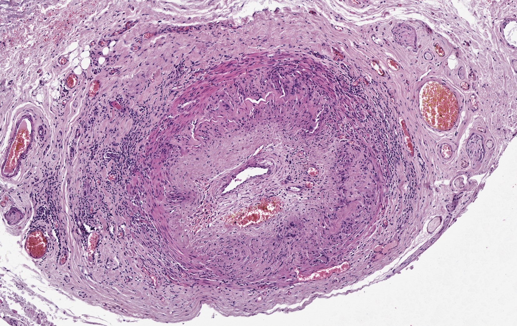
Panarteritic pattern GCA is considered when the inflammatory cells are seen distributed in all layers of the arterial wall. This pattern is the most common
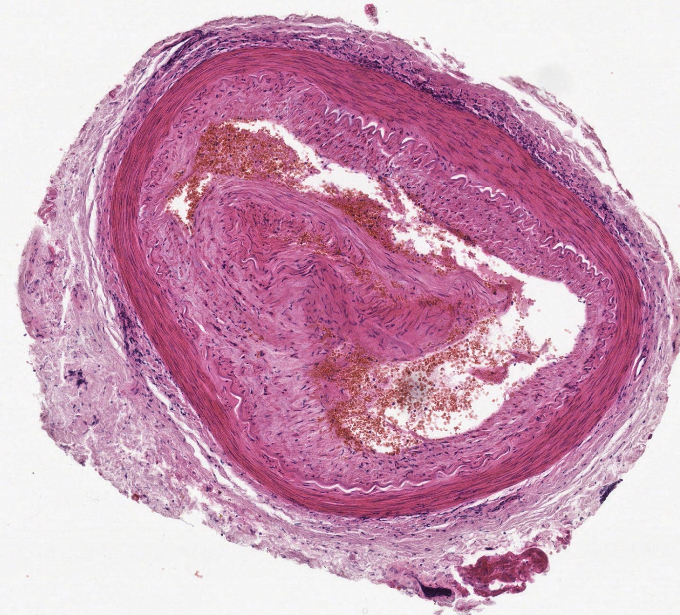
Adventitial pattern GCA is considered when inflammatory cells are restricted to the adventitia only. Please refer to visual prompt to ensure you are looking at
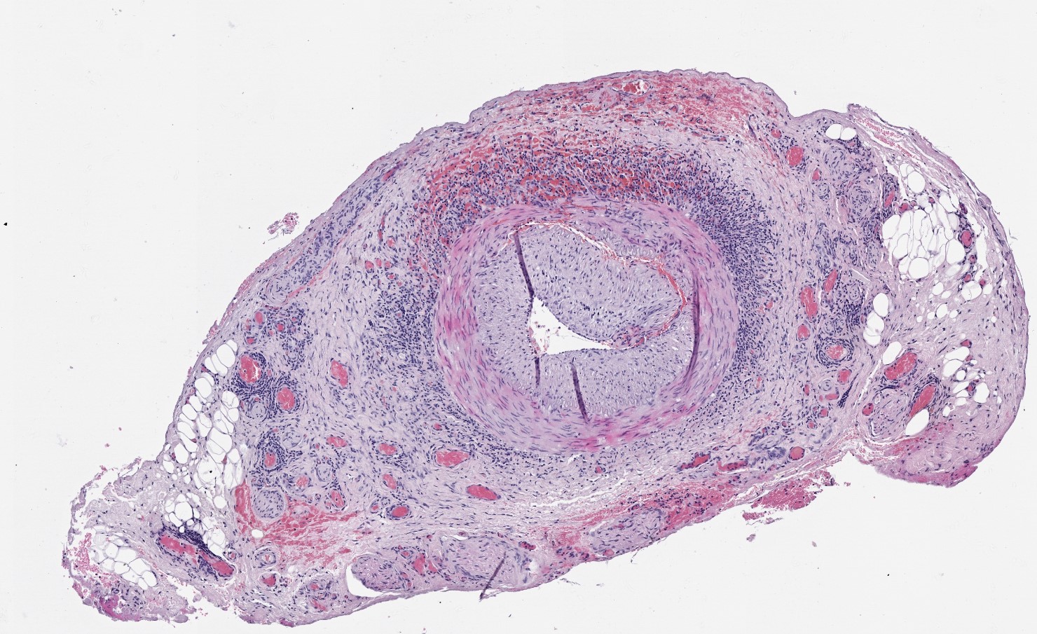
Adventitial invasive pattern GCA is considered when inflammatory cells are present in the adventitia with focal extension into the tunica media. The integrity of the
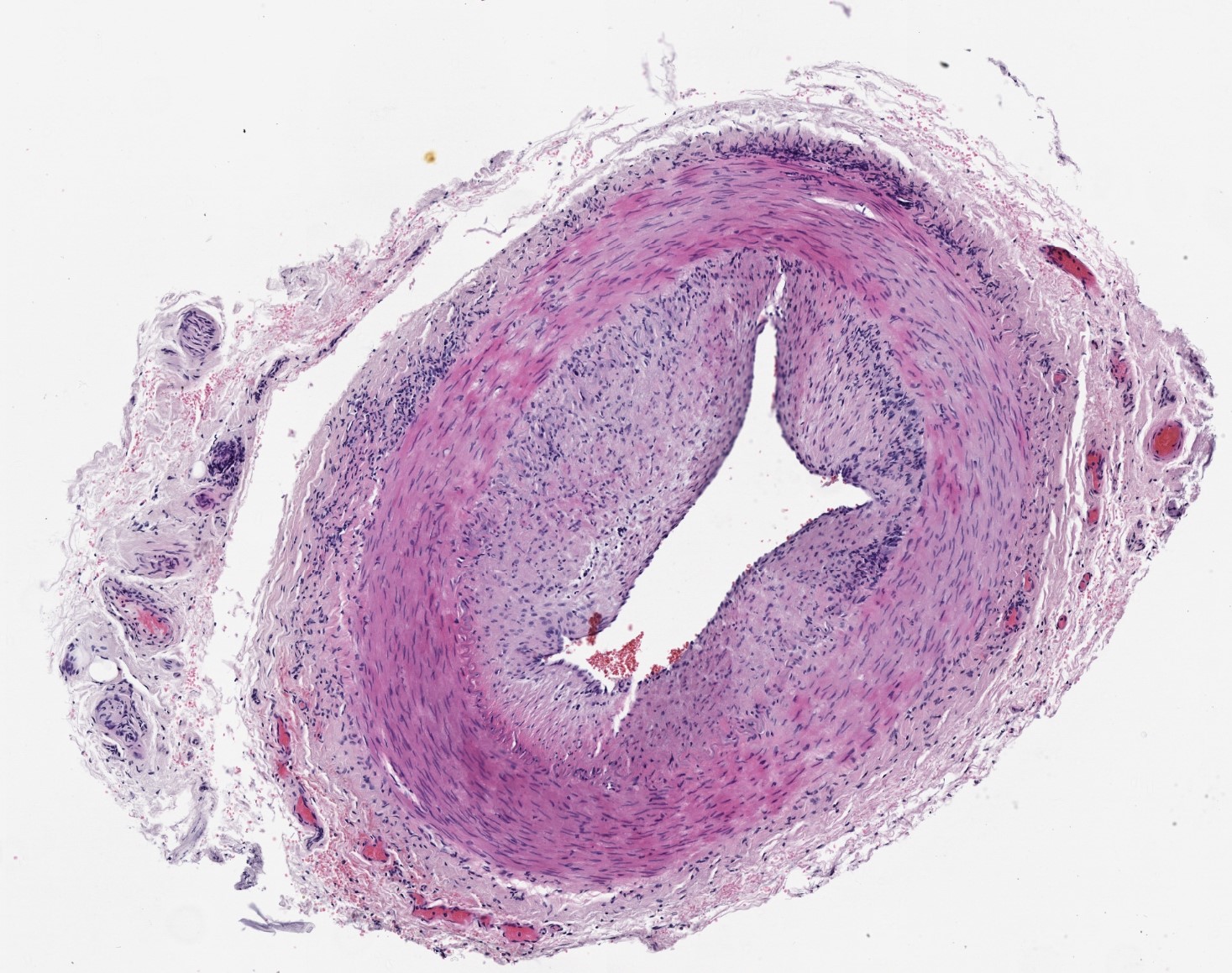
Concentric bilayer pattern GCA is considered when inflammatory cells are present in the adventitia and the tunica intima. Crucially, there is no infiltration of inflammatory


This project is funded by Leeds Hospital Charity and The Pathological Society of Great Britain and Ireland. All whole slide images are hosted on the Leeds Virtual Pathology website and we are grateful for their collaboration.
Further information on our funder and collaborators can be found here:
Leeds Hospital Charity: www.leedshospitalscharity.org.uk
The Pathological Society of Great Britain and Ireland: www.pathsoc.org
Leeds Virtual Pathology website: www.virtualpathology.leeds.ac.uk
Website developed by Proportion Marketing.