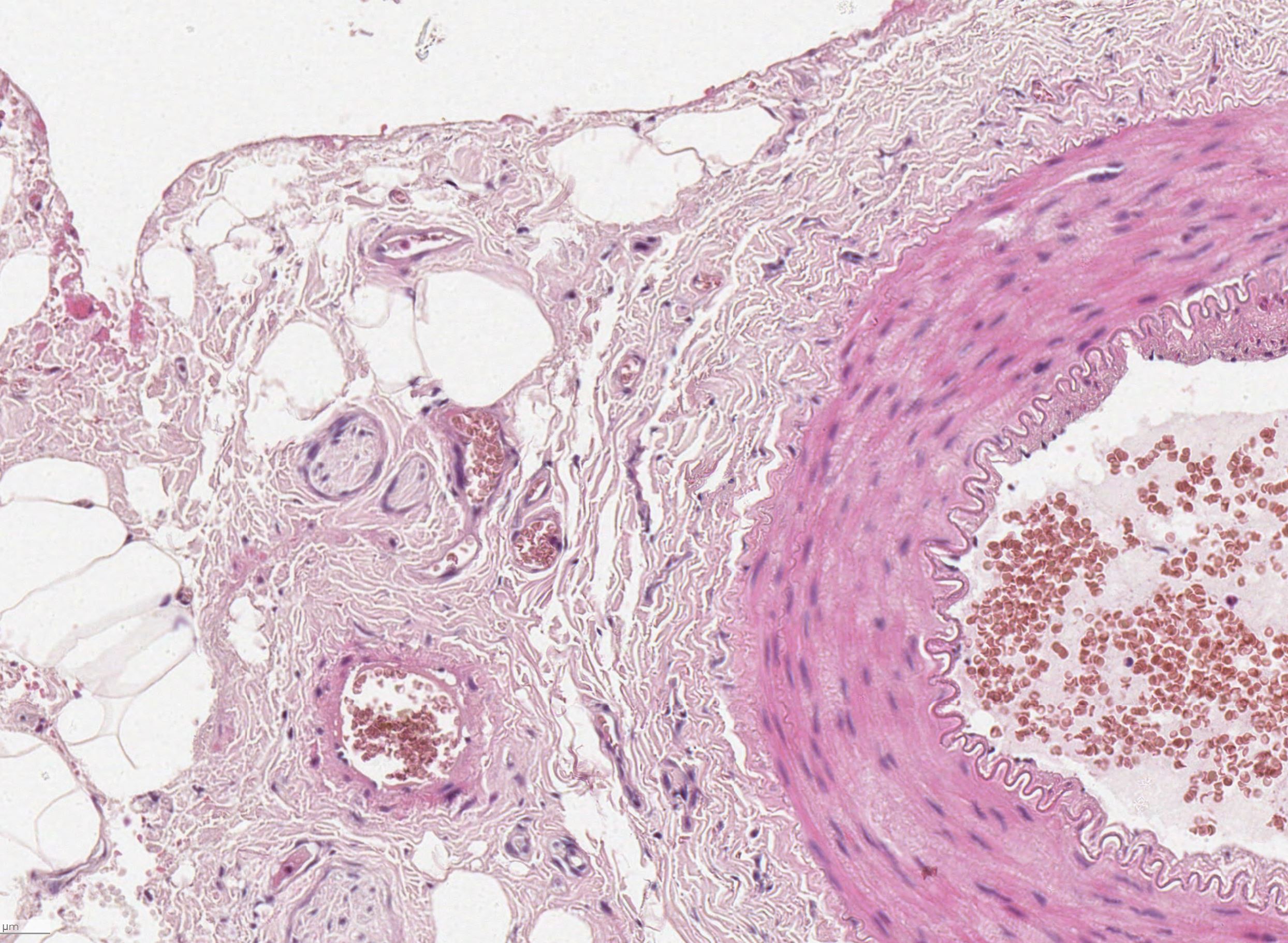
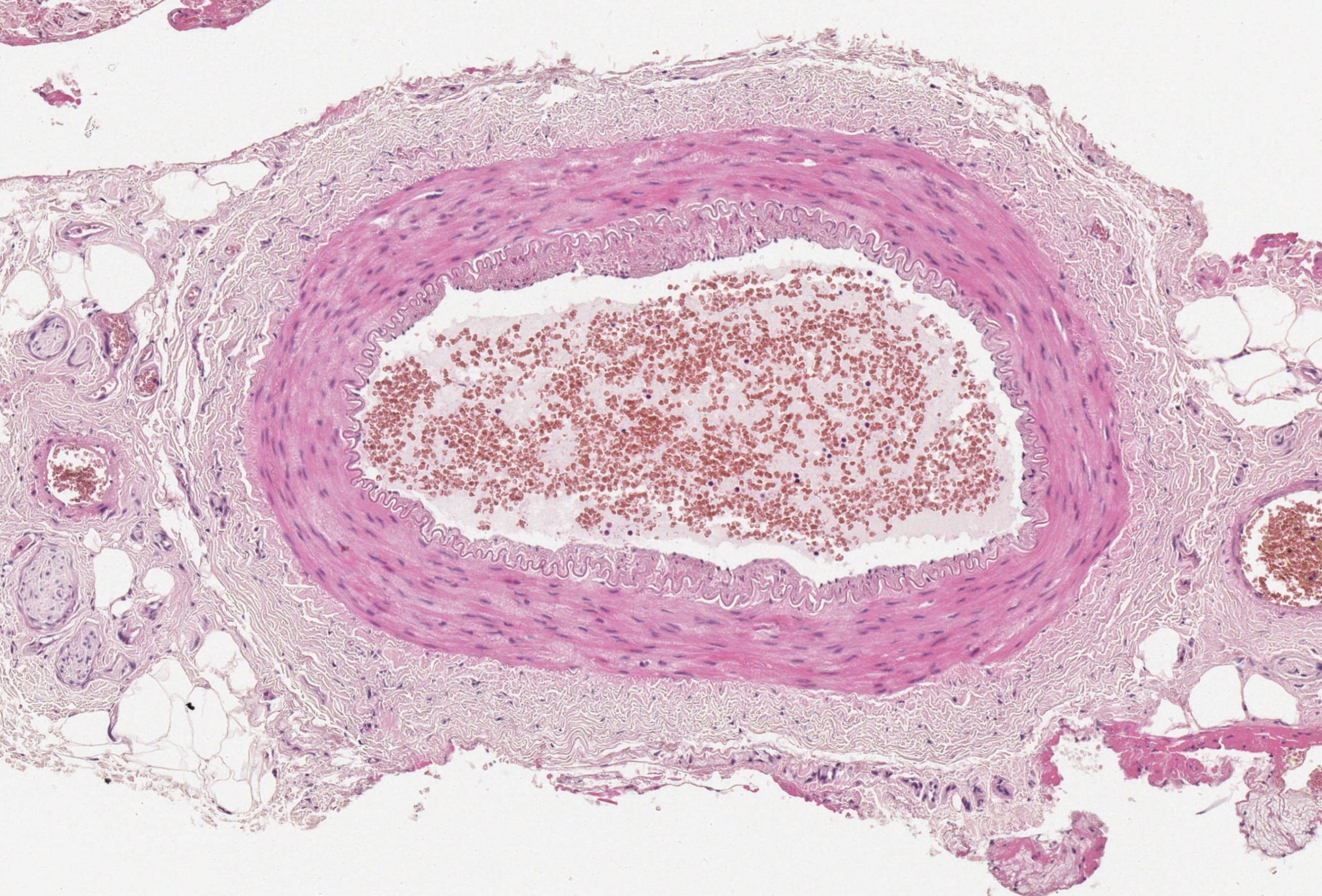
A normal temporal artery comprises 3 distinct layers. These are:
- Tunica intima
- Tunica media
- Adventitia
Each structure has its unique properties which can be summarised as follows:
| Layer | Structure |
| Tunica Intima | Comprises a single layer of endothelial cells supported by a basement membrane |
| Internal elastic lamina | Distinguishes the boundary between the intima and media |
| Tunica media | Composed of smooth muscle cells |
| Adventitia | Merges with the surrounding collagenous tissue. Contains the vasa vasorum ‘vessels of vessels’ which are a capillary network that supply oxygen and nutrients to the artery. |


